|
|
As with any medical procedure, adequate restraint is an important part of a reproductive examination. For most dairy cows, the restraint required to perform a rectal palpation is minimal. Usually a head gate or halter is sufficient. However, certain cows may require additional restraint such as a nose lead or having their tail forced firmly dorsally and cranially "jacking the tail" to decrease sudden side to side movements or kicking, both of which may cause injury to the examiner (3,4,5,9). The examiner should wear a disposable plastic sleeve or latex obstetrical sleeve, and the sleeved hand and arm should be well coated with a non-irritating, water soluble lubricant (e.g. J-jelly) (3,4,5,9). Disposable plastic sleeves may be worn inside out to keep the sleeve seam out of contact with the rectal mucosa. Some practitioners tear off the fingers of the disposable plastic sleeve and wear a latex exam glove over the hand to increase tactile sensitivity and decrease irritation to the cow's rectum (3,4). All jewelry musts be removed from the hand and arm to be inserted into the cow's rectum and the examiner's finger nails should be trimmed and clean (9).
|
|
|
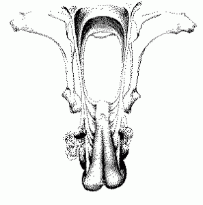
a. Pelvic cavity; b. pelvic brim; c. pelvic floor; d. pelvic walls; e. pelvic inlet; f. iliac crest; g. ilial shaft; h. ishiatic tuberosity; i. ischiatic arch.
Because palpation of the uterus per rectum relies solely on the sense of touch, it is important for the examiner to be familiar with the bony landmarks within the pelvic cavity (10). The importance of these landmarks will become clear as they are referred to in the pages to follow.
|
|
|
Placement of the gloved and lubricated hand into the rectum is achieved by holding one's hand in the shape of a cone and inserting the hand through the anus and into the rectum using a slow rotating motion with firm, but gentle pressure (3,4,9). Advancing the hand in this manner often stimulates defecation. However, additional fecal material may need to be removed from the rectum manually. This may be done by cupping the hand and gently raking feces caudally toward the anus, allowing them to pass out of the anus beside the arm without removing one's hand (4,5,9). Occasionally during an exam, the rectum may become filled with air, causing the walls of the rectum to become distended and taught. When this occurs, palpation of internal structures becomes virtually impossible. Normal peristalsis will expel this air, but the process may be expedited by reaching forward and hooking one's fingers through the perstaltic constriction band and gently pulling it caudally toward the anus, allowing air to escape beside the arm (4,9).
|
| It is common for the examiner to encounter peristaltic waves within the rectum during an examination period. These are felt as a constriction of the rectum that advances toward the anus. The hand should never be forced through one of these waves. Instead, they should be allowed to pass over the hand and arm before proceding with the examination. In small heifers, this too may be contraindicated, as even this amount of strain on rectal tissues may result in laceration (9). If fresh blood, or clotted blood in excess of 15 mls, is observed coming from the rectum during an exam, it is highly indicative of rectal rupture and requires immediate cessation of the exam and supportive therapy of the cow. Such incidences may result in sepsis, or in adhesions that can compromise reproductive function. This poses a serious health risk to the cow, and may result in a decision to cull (5, 9). In general, vigorous examination should be avoided since it not only increases the risk of harm to the cow, but also induces more peristaltic waves and rectal tone, making palpation more difficult. The examiner will find that gentleness, care and patience will reduce injury to the cow, and increase the efficiency of the procedure (3, 9). |
|
|
Once the hand and arm are in the rectum, and feces and air have been removed, the process of uterine retraction and examination may proceed. The first step is to locate the cervix. Usually the cervix lies on the midline of the floor of the pelvic cavity, but may be displaced laterally by a full bladder or a short broad ligament (9). To find the cervix, slide the hand down one wall of the pelvic cavity, and across the pelvic floor of the opposite wall, feeling for a firm, cylindrical, somewhat irregular structure lying parallel with the axis of the cow. This structure is the cervix, and should not be confused with any other structure in the pelvic cavity (10). Once located, the cervix can be grasped and, in the pregnant cow (or cow in early pregnancy), it should be freely movable. At this time the examiner should note the size, shape, form, consistency and position of the cervix (4,10). The annular folds of the cervical mucosa can be appreciated (5). Depending on the age and parity of the individual, the cervix of a normal cow can range from 5 to 12 cm in length and 2 to 6 cm in diameter, and changes little over the course of the estrous cycle (3,5). In certain breeds (e.g. Guernsey, Shorthorn), the cervix itself may extend over the pelvic brim and lie partly in the abdominal cavity (10). In the pregnant cow, the cervix becomes more enlarged (5). |
|
|
Uterine retraction is initiated by grasping the cervix and pulling it dorsally and caudally. One attempt should bring the uterine horns and broad ligament above the pelvic brim, but in some larger breeds or individuals with larger tracts, this may need to be repeated. The uterus may then be held in place by putting the thumb under the uterine body and suspending it against the pelvic wall, leaving the fingers free to locate the broad ligament (3,10).
|
|
|
While holding the uterus in place with the thumb, hook the remaining four digits around the anterior edge of the broad ligament, at the angle between the ovarian tip of the uterine horn and the ovary (10). The broad ligament, at this point, should be slightly taught as the result of the cervical retraction performed previously (3). However, it should be noted that this part of the procedure often proves to be one of the most challenging and problematic steps for the beginner (10). Hang in there!
|
|
|
With the fingers in contact with the broad ligament, run them ventrally and medially along its anterior edge to locate the uterine horn to which the ligament is attached. With short, gentle movements of the fingers, gather the horn into the hand working medially toward the uterine bifurcation. This is another step which is often difficult for the beginner who tends to lose hold of the horn (8).
|
|
|
Once the fingers have reached the base of the uterine horn, the bifurcation of the horns can be appreciated. At this time, the two intercornual ligaments can be palpated: The dorsal intercornual ligament which is the smaller and thinner of the two, and the ventral intercornual ligament which is larger and thicker. The ventral intercornual ligament will be used for retraction of the uterus, the dorsal one being too fragile for the task (3, 10). |
Ventral Intercornual Ligament
|
|
|
Hook the tip of the middle finger under the ventral intercornual ligament, and pull the uterus dorsally and caudally into the pelvic cavity, reflecting the uterus back on itself. In order for the uterus to remain in place and allow effective examination of the uterine horns, the ventral intercornual ligament should be at the level of the ischiatic arch, the cervix should be upside down, and the uterine horns should be entirely within the pelvic cavity(3,10). This procedure works well in nonpregnant cows and in cows pregnant for less than 50 - 60 days. However, in cows pregnant for more than 65 days, or who have pyometra, hydrometra, or other uterine abnormalities causing the uterus to be enlarged, heavy and possibly friable, this procedure may not be effective. Instead, such instances may require that the hand be passed beneath the uterine horns so that they may be lifted into the pelvic cavity. In later pregnancy, or in a grossly enlarged uterus, retraction is not possible (3,10). In some animals, especially nulliparous or primiparous ones, the uterus can be retracted simply by reaching forward and by directly locating the ventral intercornual ligament and reflecting the uterus caudally. This method, however, is not routine, and often fails (10). At any point during the retraction process peristaltic waves can occur and interfere with palpation, causing the examiner to lose hold of the uterus. When such peristalsis occurs, one can keep the uterus in place by cupping one's hand and pressing the uterus to the floor or wall of the pelvic cavity. Again, gentle manipulations will stimulate less peristaltic activity than vigorous palpation. As one becomes more adept, the process can be acheived in less time, which also will decrease peristaltic interference (9,10). It has been noted that wintering cows who are on dry, preserved feed have less peristaltic activity and rectal tone than cows on lush pasture in warmer seasons (5). |
|
|
Once complete retraction of the uterine horns have been acheived, a thorough examination should be performed. Starting at the base of the horn at the bifurcation and working toward the tip, examine each horn for size, form, consistency, tone and contents. It is important to be certain that the full extent of both uterine horns have been examined (3,6,8). Palpable qualities of the uterine horn change with reproductive and disease status of the cow. During a normal estrous cycle, uterine tone will begin to increase a few days before the onset of estrus, becoming fully toned at estrus. This condition will persist for about 2 days, then tone will decrease as the cow enters the luteal phase of her cycle. During the luteal phase, the horns become soft and flaccid. Therefore, it is easier to identify and palpate the uterine horns during or close to estrus (1,5). Diagnosis of pregnancy is another important function of rectal palpation. In order to diagnose a pregnancy by palpation, one must detect one or more of the "positive signs of pregnancy": 1. Fetal membrane slip, which can be appreciated from about 30 days to term; 2. amniotic vesicle, which is palpable between 30 - 65 days of pregnancy; 3. placentomes, palpable from about 75 days to term, and 4. fetus, felt from about 65 - 70 days to term (1,3). Some consider fremitus (palpable turbulance) in the uterine artery a positive sign of pregnancy, since it is rarely present in a non-pregnant individual. Fremitus is palpated during pregnancy in the uterine artery ipsilateral to the pregnant horn (3). It should be noted that prolonged and vigorous palpation of a pregnant cow can increase the risk of early embryonic death. This occurs especially as a result of attempts to palpate the amnionic vesicle and the fetal membrane slip. Fewer problems are observed with palpation of fluctuation in the uterine horn. It is best, when palpating a cow in early pregnancy, to do so gently, and recheck her after 58 -60 days for confirmation (7). Uterine size decreases quite rapidly after parturition. Normally, the uterus is small enough to allow palpation of its full extent by 7 - 10 days post-partum. It should be at normal size by about 25 days, and should be completely involuted by 42 - 46 days. The cervix returns to normal at an even faster rate (1,5). |
|
|
When thorough examination of the uterine horns is complete and the cow has been determined to be non-pregnant, or is pregnant but there is concern that there has been fetal death, one may proceed with palpation of the ovaries. Palpation of the ovaries in the normal, pregnant cow is not recommended because it may induce luteolysis and subsequent loss of the pregnancy (1,3,5,9,10). The ovary is located by finding it in relation to the tip of the uterine horn, or by recapturing the broad ligament and locating the ovary which is suspended by the mesometrium (4,10). Once the ovary is found, it is held so that the ventral "free" border faces dorsally, while the dorsal "attached" border rests in the hand, placing the mesovarium between the middle and ring fingers. The surface of the ovary is then explored using the forefinger and the thumb, allowing the examiner to evaluate ovarian size, consistency, presence of functional structures, and any abnormalities (10). Characteristics of the ovary vary between ovaries in the same individual, and between individuals, ages, and breeds. In the prepuberal heifer, the ovaries are small and smooth. In postpuberal heifers and cows, ovaries that are smaller than 2 cm in length by 1.5 cm in width by 1.5 cm in thickness can be suspected of being hypoplastic or atrophied. Normal ovaries vary in size and shape over the course of the estrous cycle, on average ranging from 2 - 4 cm long by 2 - 3 cm wide by 2 - 3 cm thick (1,5). Some increase in ovarian size is associated with developing follicles, but the greatest changes in sizes are due to the presence of the corpus luteum. If an ovary is found to be abnormally large, this may be due to a follicular or luteal cyst or to neoplastic change (10). |
|